Light from the eyes – practical Bioelectrography
Experimentally Observed Cherenkov Light Generation in the Eye During Radiation Therapy

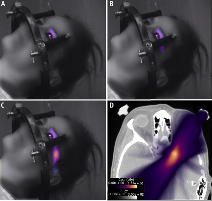
Summary: As ionizing radiation passes through the volume of the eye, light is generated within the vitreous fluid. We found that the eye acts as an integrating sphere – light created inside the eye exits through the lens. Using an intensified and time-gated camera, we captured light exiting the eye in real-time during patient and animal sample irradiation. By analyzing the spectra of this signal, we experimentally determined that it resembles Cherenkov emission. Light signal strength was largely homogeneous throughout the entire eye and the amount of light generated in the eye exceeds the visible detection threshold.
Purpose: Patients have reported sensations of seeing light flashes during radiation therapy, even with their eyes closed. These observations have been attributed to either direct excitation of retinal pigments or generation of Cherenkov light inside the eye. Both in vivo human and ex vivo animal eye imaging was used to confirm light intensity and spectra to determine its origin and overall observability.
Method and Materials: A time-gated and intensified camera was used to capture light exiting the eye of a patient undergoing stereotactic radiosurgery in real time, thereby verifying the detectability of light through the pupil. These data were compared with follow-up mechanistic imaging of ex vivo animal eyes with thin radiation beams to evaluate emission spectra and signal intensity variation with anatomic depth. Angular dependency of light emission from the eye was also measured.
Results: Patient imaging showed that light generation in the eye during radiation therapy can be captured with a signal-to-noise ratio of 68. Irradiation of ex vivo eye samples confirmed that the spectrum matched that of Cherenkov emission and that signal intensity was largely homogeneous throughout the entire eye, from the cornea to the retina, with a slight maximum near 10 mm depth. Observation of the signal external to the eye was possible through the pupil from 0° to 90°, with a detected emission near 2500 photons per millisecond (during peak emission of the ON cycle of the pulsed delivery), which is over 2 orders of magnitude higher than the visible detection threshold.
Conslusions: By quantifying the spectra and magnitude of the signal, we now have direct experimental observations that Cherenkov light is generated in the eye during radiation therapy and can contribute to perceived light flashes. Furthermore, this technique can be used to further study and measure phosphenes in the radiation therapy clinic.
International Journal of Radiation Oncology, Biology, & Physics:
https://www.redjournal.org/article/S0360-3016(19)33947-1/fulltext
Imaging of Ultraweak Spontaneous Photon Emission from Human Body Displaying Diurnal Rhythm

Abstract
The human body literally glimmers. The intensity of the light emitted by the body is 1000 times lower than the sensitivity of our naked eyes. Ultraweak photon emission is known as the energy released as light through the changes in energy metabolism. We successfully imaged the diurnal change of this ultraweak photon emission with an improved highly sensitive imaging system using cryogenic charge-coupled device (CCD) camera. We found that the human body directly and rhythmically emits light. The diurnal changes in photon emission might be linked to changes in energy metabolism.
Introduction
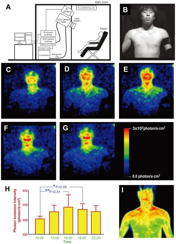
Bioluminescence, which is weak but visible, is sometimes produced in living organisms, such as fireflies or jellyfish, as the result of specialized enzymatic reactions that require adenosine triphosphate. However, virtually all living organisms emit extremely weak light, spontaneously without external photoexcitation [1]. This biophoton emission is categorized in different phenomena of light emission from bioluminescence, and is believed to be a by-product of biochemical reactions in which excited molecules are produced from bioenergetic processes that involves active oxygen species [1], [2]. Human body is glimmering with light of intensity weaker than 1/1000 times the sensitivity of naked eyes [3], [4]. By using a sensitive charge-coupled-device (CCD) camera with the ability to detect light at the level of a single photon, we succeeded in imaging the spontaneous photon emission from human bodies [3].
Previously, for obtaining an image, it took more than 1 hour of acquisition, which is practically impossible for the analysis of physiologically relevant biophoton emission. By improving the CCD camera and lens system, here we have succeeded in obtaining clear images using a short exposure time, comparable with the analysis of physiological phenomena. Since metabolic rates are known to change in a circadian fashion [5], [6], we investigated the temporal variations of biophoton emission across the day from healthy human body.
Results and Discussion
A cooled CCD camera operated at −120°C with slow scanning mode read-out was used with a specially designed high-throughput lens system. The camera was placed in a light-tight room in complete darkness (schematic illustration of the experimental setup is shown in Fig. 1A). Five healthy male volunteers, in their 20′s, were subjected to normal light-dark conditions and allowed to sleep from 0:00–7:00. On the days of photon imaging, volunteers were kept in a room (400 lux) adjacent to the dark room. For imaging purposes, the body surface was wiped and the subject was left 15 minutes in the dark room for dark adaptation, after which the naked subject in sitting position was exposed for 20 minutes to the CCD camera. Measurements were carried out in every 3 hours from 10:00 to 22:00 and continued for 3 days. Just before and after the measurements, the surface body (thermography) and oral temperature were taken. Saliva was also collected after the photon measurements for the analysis of cortisol level as a biomarker of endogenous circadian rhythms. Temporal variation of photon emission intensity was calculated from image data with extraction of the face and body intensity.
Journal of High Energe Physics
https://www.ncbi.nlm.nih.gov/pmc/articles/PMC2707605/
Cherenkov Light
Light Generation in the Eye
Naturally Light Hidden Photons in LARGE Volume String Compactifications
See more about Biophotons
The biological emission of photons (biophotons) is a term used to describe the permanent ultraweak (1-100 photons/sec/cm2) emission of coherent (phase-locked and/or frequency-locked) photons from living systems. (F.A.Popp 1976) Popp considered it to be a quantum biological phenomenon with bio-informational character distinct from the non-coherent emission of photons as by-products of metabolism, like thermal radiation and bioluminescence/chemiluminescence caused by radical reactions, oxidation etc.
Biophoton/ultraweak photon emission originates from relaxation of electronically excited states of the constituents of living cells, which are generally associated with the presence of an oxidative metabolism that accompanies the production of reactive oxygen species (ROS) which participate in the regulation of a wide spectrum of biochemical and physiological functions.
BEZ 20.02.98
Light from the eyes – an indicator of the development of civilization
Boris Evgenievich Zolotov (November 20, 1947, Bryansk Oblast – December 25, 2015, Sochi) – world-famous scientist, philosopher, actor, director, teacherю
Boris Zolotov inventor of Leader-Follow, Intuitive Information Sight, MiniMax, EOS – Expert Operator System and other the Biointernet modes, technologies and practices.
Dr. Zolotov author of 24/7 seminars (the Biointernet seminars). Since 1988, Boris Evgenievich Zolotov conducted developmental seminars in the USSR, Russia, Bulgaria, Hungary and other countries. Last years, seminars have taken the form of “miracle parties” and “meetings with interesting people.”
Light from the eyes
The Biointernet works through Human Light System

1 thought on “Light from the eyes”